


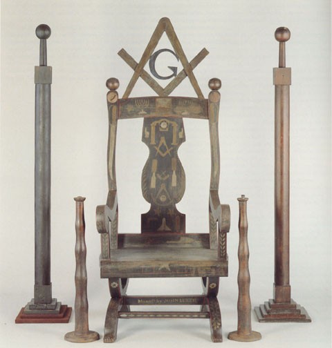
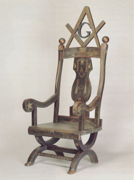
John Luker, Masonic Master’s chair, pillars, and candle stands, Vinton County, Ohio, 1870. (Courtesy, Museum of Our National Heritage; photo, David Bohl.)
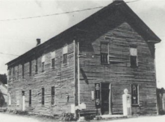
Swan Lodge No. 358, Vinton County, Ohio. This photo probably was taken during the mid-1960s. (Courtesy, Museum of Our National Heritage.)
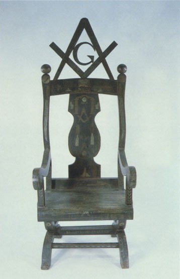
Pretreatment view of the Masonic Master’s chair illustrated in fig. 1. (Photo, Susan Buck.)
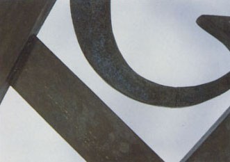
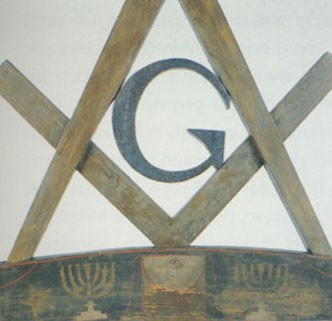
Pretreatment detail of the G, compass, and square of the Masonic Master’s chair illustrated in fig. 1. The surface of the square and dividers was corroded, and the G was encrusted with grime. (Photo, Susan Buck.)
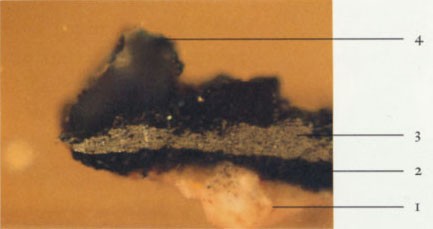
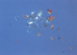
Cross-section of the G of the Masonic Master’s chair photographed in visible light at 125x: (1) white preparatory layer; (2) blue glaze layer; (3) thick pewter powder layer; (4) large smalt particle. (Photo, Susan Buck.)
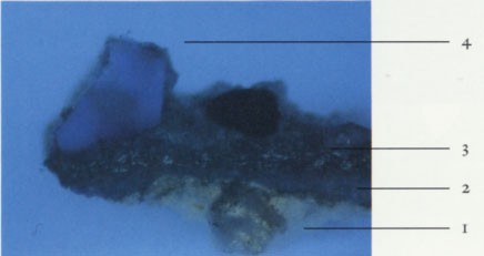
Cross-section of the G of the Masonic Master’s chair photographed in ultraviolet light at 125x: (1) white preparatory layer; (2) blue glaze layer; (3) thick pewter powder layer; (4) large smalt particle. The fluorescence indicates that the blue glaze and metallic powders have a plant resin binding medium. (Photo, Susan Buck).
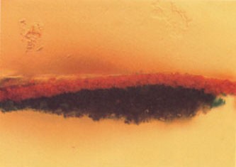
Cross-section of a red stripe on the Masonic Master’s chair photographed in visible light at 500x. (Photo, Susan Buck.)
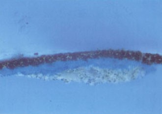
Cross-section of a red stripe on the Masonic Master’s chair photographed in ultraviolet light at 500x. The flourescence indicates that the preparatory layer, blue glaze, and red stripe have a plant resin binding medium and that there is an early plant resin varnish coating on the red stripe. (Photo, Susan Buck.)
Red lead pigment particles with characteristic bright birefringence and isotropic Prussian blue particles photographed with transmitted light under crossed polars at 500x. (Photo, Susan Buck.)
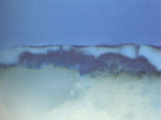
Cross-section of a protected surface under the seat of the Masonic Master’s chair photographed in ultraviolet light at 500x.
A thick layer of cracked, weathered plant resin varnish is visible above the blue glaze layer. (Photo, Susan Buck.)
Post-treatment view of the Masonic Master’s chair. The painted surfaces under the seat are closest to the original colors. (Photo, David Bohl.)
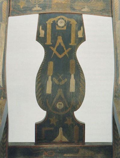
Post-treatment detail of the splat and crest rail of the Masonic Master’s chair. (Photo, David Bohl.)
Post-treatment detail of the crest rail, G, compass, and square. Although cleaning helped reveal the red paint and blue glaze, the metallic powder layers remained darkened and corroded. (Photo, David Bohl.)